Slice up the brain
Epilepsy is a severe neurological disorder characterized by recurrent epileptic seizures. These seizures result from sudden, abnormal electrical discharges in the brain, leading to temporary changes in consciousness, behavior, and perception. Shockingly, one out of three epilepsy patients continues to experience seizures despite current anti-seizure medications. Therefore, there is an imminent need for ongoing epilepsy research, such as the work conducted by a dedicated team at Ghent University.
Ever wondered how epilepsy can be studied in the laboratory? One powerful method is through hippocampal slice experiments. Let's break it down in simple terms focusing on the hippocampus and the slice experiments.
The hippocampus is a small seahorse-shaped structure deep within the brain and plays an important role in memory formation and spatial navigation. Since this structure is frequently implicated in epilepsy, studying it can offer valuable insights into the disorder's mechanisms and potential new treatments.
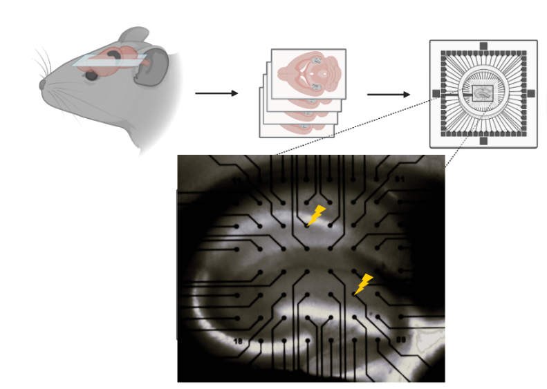
In a slice experiment, researchers start by removing the brain of a laboratory animal, such as a mouse or rat. These brains are then sliced into sections, each about 350 µm thick. Since each animal has two hippocampi, researchers can obtain a total of eight slices from one animal. These slices are carefully preserved in a specialized solution that replicates the brain's environment, allowing the neurons to remain alive and functional for up to eight hours.
In the experiment, each slice is individually tested: neurons are stimulated using electrical pulses or chemicals to simulate seizure activity. The resulting electrical activity is recorded using sophisticated equipment known as a multielectrode array, which consists of 60 tiny electrodes. This setup enables researchers to study abnormal neuron behavior and test potential new treatments.
In conclusion, hippocampal slice experiments are indispensable tools in epilepsy research. They provide a controlled laboratory environment to explore the underlying mechanisms of epilepsy and investigate potential treatments. Additionally, by maximizing the information obtained from each laboratory animal, researchers can minimize the use of animals while striving to improve the lives of those affected by this condition.
Publications: